The Organ That Contains the Penis During Sexual Intercourse and Through Which a Baby Is Born Is the
Chapter 13: Introduction to Animal Reproduction and Development
13.3 Human Reproduction
Learning Objectives
By the cease of this section, y'all will be able to:
- Describe human male and female reproductive anatomies
- Draw spermatogenesis and oogenesis and hash out their differences and similarities
- Draw the function of hormones in human reproduction
- Describe the roles of male and female person reproductive hormones
Every bit in all animals, the adaptations for reproduction in humans are complex. They involve specialized and different anatomies in the two sexes, a hormone regulation organisation, and specialized behaviors regulated past the encephalon and endocrine organization.
Human Reproductive Anatomy
The reproductive tissues of male person and female humans develop similarly in utero until well-nigh the seventh week of gestation when a low level of the hormone testosterone is released from the gonads of the developing male person. Testosterone causes the primitive gonads to differentiate into male sexual organs. When testosterone is absent, the primitive gonads develop into ovaries. Tissues that produce a penis in males produce a clitoris in females. The tissue that will become the scrotum in a male becomes the labia in a female. Thus the male and female anatomies arise from a departure in the development of what were once mutual embryonic structures.
Male person Reproductive Anatomy
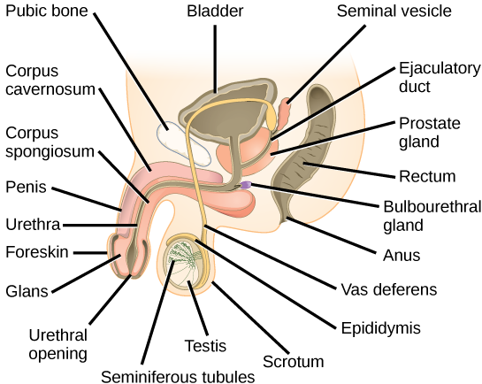
Sperm are immobile at body temperature; therefore, the testes are external to the body so that a correct temperature is maintained for movement. In land mammals, including humans, the pair of testes must be suspended exterior the body so the environment of the sperm is about two °C lower than body temperature to produce feasible sperm. If the testes exercise not descend through the abdominal cavity during fetal development, the individual has reduced fertility.
The scrotum houses the testicles or testes (singular: testis), and provides passage for blood vessels, nerves, and muscles related to testicular function. The testes are a pair of male gonads that produce sperm and reproductive hormones. Each testis is approximately ii.five by iii.viii cm (1.5 by ane inch) in size and divided into wedge-shaped lobes past septa. Coiled in each wedge are seminiferous tubules that produce sperm.
The penis drains urine from the urinary float and is a copulatory organ during intercourse (Effigy 13.12; Table thirteen.1). The penis contains three tubes of erectile tissue that become engorged with blood, making the penis cock, in preparation for intercourse. The organ is inserted into the vagina culminating with an ejaculation. During orgasm, the accessory organs and glands continued to the testes contract and empty the semen (containing sperm) into the urethra and the fluid is expelled from the body by muscular contractions causing ejaculation. After intercourse, the blood drains from the erectile tissue and the penis becomes flaccid.
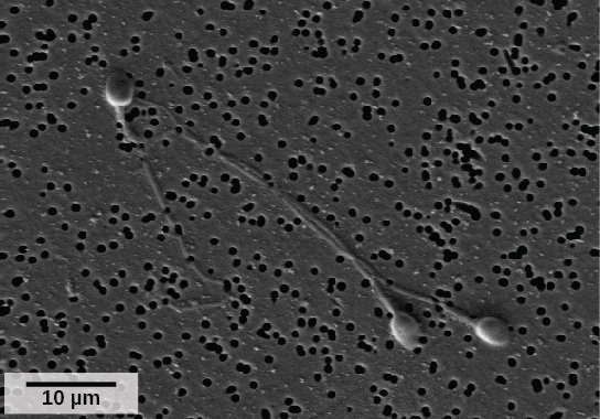
Semen is a mixture of sperm (nigh 5 percentage of the total) and fluids from accompaniment glands that contribute most of the semen'southward volume. Sperm are haploid cells, consisting of a flagellum for motility, a neck that contains the cell's energy-producing mitochondria, and a caput that contains the genetic material (Figure thirteen.11). An acrosome (acrosomal vesicle) is institute at the top of the head of the sperm. This construction contains enzymes that can assimilate the protective coverings that surround the egg and allow the sperm to fuse with the egg. An ejaculate will contain from ii to five milliliters of fluid and from 50–120 million sperm per milliliter.
Sperm form in the walls of seminiferous tubules that are coiled inside the testes (Figure xiii.12; Tabular array xiii.one). The walls of the seminiferous tubules are made upwardly of the developing sperm cells, with the to the lowest degree developed sperm at the periphery of the tubule and the fully developed sperm side by side to the lumen. The sperm cells are associated with Sertoli cells that nourish and promote the development of the sperm. Other cells present between the walls of the tubules are the interstitial cells of Leydig, which produce testosterone once the male reaches boyhood.
When the sperm have developed flagella they leave the seminiferous tubules and enter the epididymis (Effigy xiii.12; Table thirteen.ane). This construction lies along the peak and posterior of the testes and is the site of sperm maturation. The sperm leave the epididymis and enter the vas deferens, which carries the sperm backside the bladder, and forms the ejaculatory duct with the duct from the seminal vesicles. During a vasectomy, a section of the vas deferens is removed, preventing sperm (but not the secretions of the accompaniment glands) from being passed out of the trunk during ejaculation and preventing fertilization.
The bulk of the semen comes from the accessory glands associated with the male reproductive system. These are the seminal vesicles, the prostate gland, and the bulbourethral gland (Figure thirteen.12; Table 13.one). The secretions from the accessory glands provide important compounds for the sperm including nutrients, electrolytes, and pH buffering. In that location are likewise coagulation factors that touch sperm delivery and motility.
Which of the following statements about the male reproductive system is faux?
A. The vas deferens carries sperm from the testes to the seminal vesicles.
B. The ejaculatory duct joins the urethra.
C. Both the prostate and the bulbourethral glands produce components of the semen.
D. The prostate gland is located in the testes.
<!–D–>
| Organ | Location | Role |
|---|---|---|
| Scrotum | External | Supports testes and regulates their temperature |
| Penis | External | Delivers urine, copulating organ |
| Testes | Internal | Produce sperm and male hormones |
| Seminal Vesicles | Internal | Contribute to semen production |
| Prostate Gland | Internal | Contributes to semen production |
| Bulbourethtral Glands | Internal | Neutralize urine in urethra |
Female Reproductive Anatomy
The breasts consist of mammary glands and fatty. Each gland consists of 15 to 25 lobes that take ducts that empty at the nipple and that supply the nursing kid with nutrient- and antibody-rich milk to aid development and protect the child.
Internal female reproductive structures include ovaries, oviducts, the uterus, and the vagina (Effigy 13.thirteen; Table 13.2). The pair of ovaries is held in identify in the intestinal crenel past a system of ligaments. The outermost layer of the ovary is made up of follicles, each consisting of 1 or more follicular cells that surroundings, nourish, and protect a single egg. During the menstrual menses, a batch of follicular cells develops and prepares their eggs for release. At ovulation, one follicle ruptures and one egg is released. Post-obit ovulation, the follicular tissue that surrounded the ovulated egg stays within the ovary and grows to course a solid mass chosen the corpus luteum. The corpus luteum secretes additional estrogen and the hormone progesterone that helps maintain the uterine lining during pregnancy. The ovaries also produce hormones, such equally estrogen.
The oviducts, or fallopian tubes, extend from the uterus in the lower intestinal cavity to the ovaries, just they are not in contact with the ovaries. The lateral ends of the oviducts flare out into a trumpet-like structure and have a fringe of finger-like projections called fimbrae. When an egg is released at ovulation, the fimbrae help the nonmotile egg enter into the tube. The walls of the oviducts accept a ciliated epithelium over shine muscle. The cilia beat, and the polish muscle contracts, moving the egg toward the uterus. Fertilization commonly takes place within the oviduct and the developing embryo is moved toward the uterus. It usually takes the egg or embryo a week to travel through the oviduct.
Sterilization in women is called a tubal ligation; information technology is analogous to a vasectomy in males in that the oviducts are severed and sealed, preventing sperm from reaching the egg.
The uterus is a structure nearly the size of a woman's fist. The uterus has a thick muscular wall and is lined with an endometrium rich in blood vessels and fungus glands that develop and thicken during the female cycle. Thickening of the endometrium prepares the uterus to receive the fertilized egg or zygote, which volition and then implant itself in the endometrium. The uterus supports the developing embryo and fetus during gestation. Contractions of the smooth muscle in the uterus help in forcing the baby through the vagina during labor. If fertilization does not occur, a portion of the lining of the uterus sloughs off during each menstrual period. The endometrium builds up once more in preparation for implantation. Part of the uterus, chosen the cervix, protrudes into the elevation of the vagina.
The vagina is a muscular tube that serves several purposes. Information technology allows menstrual catamenia to go out the torso. Information technology is the receptacle for the penis during intercourse and the pathway for the delivery of offspring.
| Organ | Location | Function |
|---|---|---|
| Clitoris | External | Sensory organ |
| Mons pubis | External | Fat area overlying pubic bone |
| Labia majora | External | Covers labia minora; contains sweat and sebaceous glands |
| Labia minora | External | Covers entrance hall |
| Greater vestibular glands | External | Secrete mucus; lubricate vagina |
| Breast | External | Produces and delivers milk |
| Ovaries | Internal | Produce and develop eggs |
| Oviducts | Internal | Transport egg to uterus; site of fertilization |
| Uterus | Internal | Supports developing embryo |
| Vagina | Internal | Common tube for intercourse, nativity culvert, passing menstrual catamenia |
Gametogenesis (Spermatogenesis and Oogenesis)
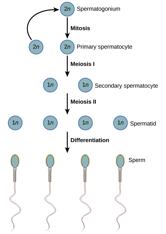
Gametogenesis, the product of sperm and eggs, involves the process of meiosis. During meiosis, ii nuclear divisions separate the paired chromosomes in the nucleus then split up the chromatids that were fabricated during an earlier phase of the cell'southward life cycle. Meiosis and its associated cell divisions produces haploid cells with half of each pair of chromosomes unremarkably institute in diploid cells. The production of sperm is called spermatogenesis and the product of eggs is called oogenesis.
Spermatogenesis
Spermatogenesis occurs in the wall of the seminiferous tubules, with the almost archaic cells at the periphery of the tube and the near mature sperm at the lumen of the tube (Figure 13.14). Immediately under the capsule of the tubule are diploid, undifferentiated cells. These stem cells, each chosen a spermatogonium (pl. spermatogonia), go through mitosis to produce one cell that remains as a stem jail cell and a second cell called a primary spermatocyte that volition undergo meiosis to produce sperm.
The diploid primary spermatocyte goes through meiosis I to produce two haploid cells chosen secondary spermatocytes. Each secondary spermatocyte divides after meiosis 2 to produce two cells called spermatids. The spermatids eventually accomplish the lumen of the tubule and abound a flagellum, condign sperm cells. Four sperm result from each primary spermatocyte that goes through meiosis.
Concept in Activeness

Visit this site to see the procedure of spermatogenesis.
Oogenesis
Oogenesis occurs in the outermost layers of the ovaries. As with sperm production, oogenesis starts with a germ cell. In oogenesis, this germ cell is called an oogonium and forms during the embryological development of the individual. The oogonium undergoes mitosis to produce about one to two million oocytes by the time of birth.
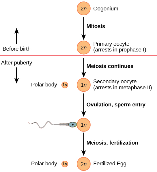
The chief oocytes brainstorm meiosis before nascency (Figure xiii.15). All the same, the meiotic segmentation is arrested in its progress in the commencement prophase stage. At the time of birth, all future eggs are in prophase I. This state of affairs is in dissimilarity with the male reproductive system in which sperm are produced continuously throughout the life of the private. Starting at adolescence, anterior pituitary hormones crusade the development of a few follicles in an ovary each month. This results in a primary oocyte finishing the beginning meiotic sectionalisation. The jail cell divides unequally, with well-nigh of the cytoplasm and organelles going to one prison cell, called a secondary oocyte, and only one set of chromosomes and a small-scale amount of cytoplasm going to the other prison cell. This second cell is chosen a polar body and usually dies. Prison cell division is over again arrested, this time at metaphase Two. At ovulation, this secondary oocyte is released and travels toward the uterus through the oviduct. If the secondary oocyte is fertilized, the cell continues through meiosis II, producing a second polar body and haploid egg, which fuses with the haploid sperm to form a fertilized egg (zygote) containing all 46 chromosomes.
Hormonal Control of Reproduction
The human male person and female reproductive cycles are controlled by the interaction of hormones from the hypothalamus and anterior pituitary with hormones from reproductive tissues and organs. In both sexes, the hypothalamus monitors and causes the release of hormones from the inductive pituitary gland. When the reproductive hormone is required, the hypothalamus sends a gonadotropin-releasing hormone (GnRH) to the anterior pituitary. This causes the release of follicle stimulating hormone (FSH) and luteinizing hormone (LH) from the anterior pituitary into the blood. Although these hormones are named later on their functions in female reproduction, they are produced in both sexes and play important roles in controlling reproduction. Other hormones have specific functions in the male person and female reproductive systems.
Male Hormones
At the onset of puberty, the hypothalamus causes the release of FSH and LH into the male system for the first fourth dimension. FSH enters the testes and stimulates the Sertoli cells located in the walls of the seminiferous tubules to begin promoting spermatogenesis (Effigy 13.16). LH also enters the testes and stimulates the interstitial cells of Leydig, located in between the walls of the seminiferous tubules, to brand and release testosterone into the testes and the blood.
Testosterone stimulates spermatogenesis. This hormone is also responsible for the secondary sexual characteristics that develop in the male during boyhood. The secondary sexual practice characteristics in males include a deepening of the voice, the growth of facial, axillary, and pubic hair, an increase in muscle bulk, and the beginnings of the sex drive.
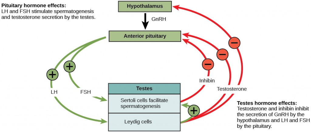
A negative feedback system occurs in the male with rising levels of testosterone acting on the hypothalamus and anterior pituitary to inhibit the release of GnRH, FSH, and LH. In addition, the Sertoli cells produce the hormone inhibin, which is released into the blood when the sperm count is likewise high. This inhibits the release of GnRH and FSH, which will cause spermatogenesis to boring down. If the sperm count reaches a low of 20 million/mL, the Sertoli cells cease the release of inhibin, and the sperm count increases.
Female Hormones
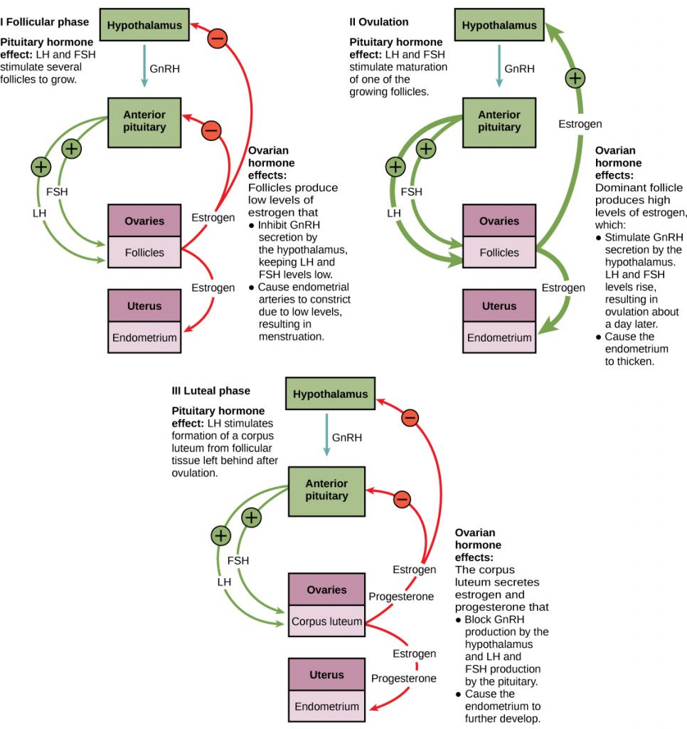
The control of reproduction in females is more than complex. The female reproductive cycle is divided into the ovarian bike and the menstrual bicycle. The ovarian cycle governs the training of endocrine tissues and release of eggs, while the menstrual bicycle governs the grooming and maintenance of the uterine lining (Figure xiii.17). These cycles are coordinated over a 22–32 day cycle, with an average length of 28 days.
As with the male, the GnRH from the hypothalamus causes the release of the hormones FSH and LH from the inductive pituitary. In add-on, estrogen and progesterone are released from the developing follicles. Equally with testosterone in males, estrogen is responsible for the secondary sexual characteristics of females. These include breast development, flaring of the hips, and a shorter period for bone growth.
The Ovarian Cycle and the Menstrual Cycle
The ovarian and menstrual cycles are regulated past hormones of the hypothalamus, pituitary, and ovaries (Effigy 13.17). The ebb and flow of the hormones causes the ovarian and menstrual cycles to advance. The ovarian and menstrual cycles occur concurrently. The commencement half of the ovarian cycle is the follicular phase. Slowly rising levels of FSH crusade the growth of follicles on the surface of the ovary. This process prepares the egg for ovulation. As the follicles grow, they begin releasing estrogen. The first few days of this cycle coincide with flow or the sloughing off of the functional layer of the endometrium in the uterus. Afterward nigh five days, estrogen levels rise and the menstrual cycle enters the proliferative stage. The endometrium begins to regrow, replacing the blood vessels and glands that deteriorated during the end of the concluding bicycle.
Which of the following statements almost hormone regulation of the female reproductive cycle is fake?
A. LH and FSH are produced in the pituitary, and estrogen and progesterone are produced in the ovaries.
B. Estradiol and progesterone secreted from the corpus luteum cause the endometrium to thicken.
C. Both progesterone and estrogen are produced by the follicles.
D. Secretion of GnRH by the hypothalamus is inhibited by low levels of estrogen but stimulated by loftier levels of estrogen.
<!– C–>
But prior to the middle of the cycle (approximately day fourteen), the high level of estrogen causes FSH and peculiarly LH to rise speedily so autumn. The spike in LH causes the almost mature follicle to rupture and release its egg. This is ovulation. The follicles that did non rupture degenerate and their eggs are lost. The level of estrogen decreases when the actress follicles degenerate.
Following ovulation, the ovarian cycle enters its luteal phase and the menstrual cycle enters its secretory stage, both of which run from virtually day 15 to 28. The luteal and secretory phases refer to changes in the ruptured follicle. The cells in the follicle undergo concrete changes and produce a construction chosen a corpus luteum. The corpus luteum produces estrogen and progesterone. The progesterone facilitates the regrowth of the uterine lining and inhibits the release of further FSH and LH. The uterus is being prepared to accept a fertilized egg, should it occur during this cycle. The inhibition of FSH and LH prevents whatsoever farther eggs and follicles from developing, while the progesterone is elevated. The level of estrogen produced by the corpus luteum increases to a steady level for the side by side few days.
If no fertilized egg is implanted into the uterus, the corpus luteum degenerates and the levels of estrogen and progesterone decrease. The endometrium begins to degenerate as the progesterone levels drib, initiating the next menstrual cycle. The decrease in progesterone also allows the hypothalamus to transport GnRH to the anterior pituitary, releasing FSH and LH and starting the cycles over again.
Reproductive Endocrinologist
A reproductive endocrinologist is a md who treats a multifariousness of hormonal disorders related to reproduction and infertility in both men and women. The disorders include menstrual problems, infertility, pregnancy loss, sexual dysfunction, and menopause. Doctors may use fertility drugs, surgery, or assisted reproductive techniques (Art) in their therapy. ART involves the utilise of procedures to dispense the egg or sperm to facilitate reproduction, such equally in vitro fertilization.
Reproductive endocrinologists undergo extensive medical training, offset in a 4-yr residency in obstetrics and gynecology, and so in a iii-year fellowship in reproductive endocrinology. To be board certified in this area, the medico must pass written and oral exams in both areas.
Gestation
Pregnancy begins with the fertilization of an egg and continues through to the birth of the private. The length of time of gestation, or the gestation menses, in humans is 266 days and is like in other great apes.
Within 24 hours of fertilization, the egg nucleus has finished meiosis and the egg and sperm nuclei fuse. With fusion, the cell is known as a zygote. The zygote initiates cleavage and the developing embryo travels through the oviduct to the uterus. The developing embryo must implant into the wall of the uterus within seven days, or it will deteriorate and die. The outer layers of the developing embryo or blastocyst grow into the endometrium past digesting the endometrial cells, and healing of the endometrium closes up the blastocyst into the tissue. Another layer of the blastocyst, the chorion, begins releasing a hormone called man beta chorionic gonadotropin (β-HCG), which makes its way to the corpus luteum and keeps that structure active. This ensures adequate levels of progesterone that will maintain the endometrium of the uterus for the support of the developing embryo. Pregnancy tests determine the level of β-HCG in urine or serum. If the hormone is present, the examination is positive.
The gestation menses is divided into three equal periods or trimesters. During the first two-to-four weeks of the showtime trimester, diet and waste matter are handled by the endometrial lining through diffusion. Every bit the trimester progresses, the outer layer of the embryo begins to merge with the endometrium, and the placenta forms. The placenta takes over the nutrient and waste requirements of the embryo and fetus, with the mother's blood passing nutrients to the placenta and removing waste from it. Chemicals from the fetus, such as bilirubin, are processed by the female parent's liver for elimination. Some of the mother'due south immunoglobulins will pass through the placenta, providing passive immunity confronting some potential infections.
Internal organs and body structures begin to develop during the first trimester. By five weeks, limb buds, optics, the heart, and liver have been basically formed. By 8 weeks, the term fetus applies, and the torso is essentially formed (Effigy thirteen.18a). The individual is well-nigh five centimeters (ii inches) in length and many of the organs, such every bit the lungs and liver, are non yet operation. Exposure to any toxins is specially dangerous during the showtime trimester, as all of the trunk's organs and structures are going through initial development. Annihilation that interferes with chemical signaling during that development tin can have a severe effect on the fetus' survival.
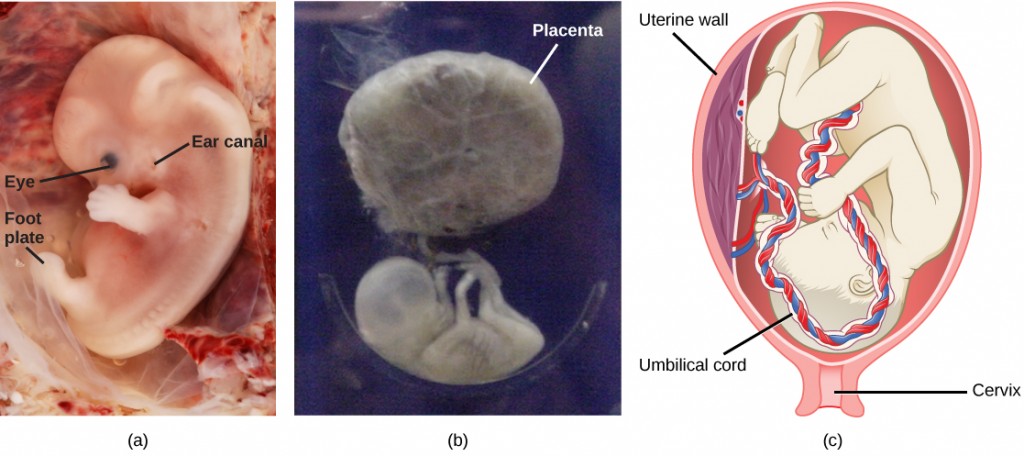
During the 2nd trimester, the fetus grows to about xxx cm (about 12 inches) (Figure 13.18b). It becomes active and the mother normally feels the first movements. All organs and structures continue to develop. The placenta has taken over the functions of diet and waste elimination and the production of estrogen and progesterone from the corpus luteum, which has degenerated. The placenta volition proceed operation up through the delivery of the baby. During the third trimester, the fetus grows to 3 to 4 kg (half dozen.5–8.v lbs.) and about 50 cm (19–xx inches) long (Effigy 13.eighteenc). This is the period of the most rapid growth during the pregnancy as all organ systems go on to grow and develop.
Concept in Action

Visit this website to see the stages of human fetal evolution.
Labor is the muscular contractions to expel the fetus and placenta from the uterus. Toward the end of the third trimester, estrogen causes receptors on the uterine wall to develop and bind the hormone oxytocin. At this time, the baby reorients, facing forward and down with the dorsum or crown of the head engaging the cervix (uterine opening). This causes the cervix to stretch and nerve impulses are sent to the hypothalamus, which signals the release of oxytocin from the posterior pituitary. Oxytocin causes shine muscle in the uterine wall to contract. At the aforementioned time, the placenta releases prostaglandins into the uterus, increasing the contractions. A positive feedback relay occurs between the uterus, hypothalamus, and the posterior pituitary to assure an adequate supply of oxytocin. As more than smooth muscle cells are recruited, the contractions increase in intensity and strength.
There are three stages to labor. During phase ane, the cervix thins and dilates. This is necessary for the baby and placenta to exist expelled during birth. The cervix volition eventually dilate to about 10 cm. During stage two, the babe is expelled from the uterus. The uterus contracts and the mother pushes as she compresses her intestinal muscles to assist the delivery. The last phase is the passage of the placenta later on the babe has been born and the organ has completely disengaged from the uterine wall. If labor should stop earlier stage 2 is reached, synthetic oxytocin, known as Pitocin, can be administered to restart and maintain labor.
Section Summary
The reproductive structures that evolved in country animals allow males and females to mate, fertilize internally, and support the growth and evolution of offspring. Gametogenesis, the production of sperm (spermatogenesis) and eggs (oogenesis), takes identify through the process of meiosis.
The male person and female reproductive cycles are controlled by hormones released from the hypothalamus and anterior pituitary and hormones from reproductive tissues and organs. The hypothalamus monitors the need for FSH and LH production and release from the anterior pituitary. FSH and LH bear on reproductive structures to cause the germination of sperm and the preparation of eggs for release and possible fertilization. In the male, FSH and LH stimulate Sertoli cells and interstitial cells of Leydig in the testes to facilitate sperm product. The Leydig cells produce testosterone, which also is responsible for the secondary sexual characteristics of males. In females, FSH and LH crusade estrogen and progesterone to be produced. They regulate the female reproductive cycle, which is divided into the ovarian cycle and the menstrual wheel.
Man pregnancy begins with fertilization of an egg and gain through the three trimesters of gestation. The starting time trimester lays down the bones structures of the trunk, including the limb buds, heart, eyes, and the liver. The 2nd trimester continues the evolution of all of the organs and systems. The tertiary trimester exhibits the greatest growth of the fetus and culminates in labor and commitment. The labor process has three stages (contractions, delivery of the fetus, and expulsion of the placenta), each propelled by hormones.
Exercises
- Which of the post-obit statements well-nigh the male reproductive arrangement is false?
- The vas deferens carries sperm from the testes to the seminal vesicles.
- The ejaculatory duct joins the urethra.
- Both the prostate and the bulbourethral glands produce components of the semen.
- The prostate gland is located in the testes.
- Which of the post-obit statements about hormone regulation of the female person reproductive cycle is faux?
- LH and FSH are produced in the pituitary, and estrogen and progesterone are produced in the ovaries.
- Estradiol and progesterone secreted from the corpus luteum cause the endometrium to thicken.
- Both progesterone and estrogen are produced by the follicles.
- Secretion of GnRH by the hypothalamus is inhibited by low levels of estrogen merely stimulated past loftier levels of estrogen.
- Sperm are produced in the ________.
- scrotum
- seminal vesicles
- seminiferous tubules
- prostate gland
- Which female person organ has an endometrial lining that will back up a developing babe?
- labia minora
- chest
- ovaries
- uterus
- Which hormone causes FSH and LH to be released?
- testosterone
- estrogen
- GnRH
- progesterone
- Nutrient and waste requirements for the developing fetus are handled during the commencement few weeks past ________.
- the placenta
- improvidence through the endometrium
- the chorion
- the blastocyst
- Which hormone is primarily responsible for the contractions during labor?
- oxytocin
- estrogen
- β-HCG
- progesterone
- Compare spermatogenesis and oogenesis equally to timing of the processes, and the number and type of cells finally produced.
- Draw the events in the ovarian cycle leading up to ovulation.
- Describe the stages of labor.
Answers
- D
- C
- C
- D
- C
- B
- A
- Stem cells are laid down in the male during gestation and lie dormant until adolescence. Stem cells in the female increase to one to ii million and enter the first meiotic partition and are arrested in prophase. At adolescence, spermatogenesis begins and continues until death, producing the maximum number of sperm with each meiotic division. Oogenesis continues again at adolescence in batches of eggs with each menstrual cycle. These primary oocytes cease the showtime meiotic division, producing a viable egg with about of the cytoplasm and its contents, and a 2nd cell chosen a polar trunk containing 23 chromosomes. The second meiotic division is initiated and arrested in metaphase. At ovulation, one egg is released. If this egg is fertilized, it finishes the second meiotic division. This is a diploid, fertilized egg.
- Low levels of progesterone permit the hypothalamus to send GnRH to the anterior pituitary and crusade the release of FSH and LH. FSH stimulates follicles on the ovary to grow and prepare the eggs for ovulation. Equally the follicles increase in size, they brainstorm to release estrogen and a low level of progesterone into the claret. The level of estrogen rises to a peak, causing a spike in the concentration of LH. This causes the most mature follicle to rupture and ovulation occurs.
- Stage 1 of labor results in uterine contractions, which thin the cervix and amplify the cervical opening. Stage ii delivers the baby, and stage three delivers the placenta.
Glossary
bulbourethral gland: the paired glands in the human male that produce a secretion that cleanses the urethra prior to ejaculation
corpus luteum: the endocrine tissue that develops from an ovarian follicle after ovulation; secretes progesterone and estrogen during pregnancy
clitoris: a sensory and erectile structure in female mammals, homologous to the male penis, stimulated during sexual arousal
estrogen: a reproductive hormone in females that assists in endometrial regrowth, ovulation, and calcium absorption
follicle stimulating hormone (FSH): a reproductive hormone that causes sperm production in men and follicle development in women
gestation: the development before nascency of a viviparous animal
gestation catamenia: the length of time of evolution, from conception to birth, of the young of a viviparous animal
gonadotropin-releasing hormone (GnRH): a hormone from the hypothalamus that causes the release of FSH and LH from the anterior pituitary
human being beta chorionic gonadotropin (β-HCG): a hormone produced by the chorion of the zygote that helps to maintain the corpus luteum and elevated levels of progesterone
inhibin: a hormone made by Sertoli cells, provides negative feedback to hypothalamus in command of FSH and GnRH release
interstitial cell of Leydig: a cell type constitute next to the seminiferous tubules that makes testosterone
labia majora: the large folds of tissue covering inguinal expanse
labia minora: the smaller folds of tissue within labia majora
luteinizing hormone (LH): a reproductive hormone in both men and women, causes testosterone product in men and ovulation and lactation in women
menstrual bike: the cycle of the degradation and re-growth of the endometrium
oogenesis: the process of producing haploid eggs
ovarian cycle: the cycle of preparation of egg for ovulation and the conversion of the follicle to the corpus luteum
oviduct: (also, fallopian tube) the muscular tube connecting uterus with ovary area
ovulation: the release of an oocyte from a mature follicle in the ovary of a vertebrate
penis: the male person reproductive structure for urine elimination and copulation
placenta: the organ that supports the transport of nutrients and waste between the mothers and fetus' blood in eutherian mammals
progesterone: a reproductive hormone in women; assists in endometrial regrowth and inhibition of FSH and LH release
prostate gland: a structure that is a mixture of smooth muscle and glandular material and that contributes to semen
scrotum: a sac containing testes, exterior to trunk
semen: a fluid mixture of sperm and supporting materials
seminal vesicle: a secretory accessory gland in male; contributes to semen
seminiferous tubule: the structures within which sperm production occurs in the testes
Sertoli cell: a cell in the walls of the seminiferous tubules that assists developing sperm and secretes inhibin
spermatogenesis: the procedure of producing haploid sperm
testes: a pair of male reproductive organs
testosterone: a reproductive hormone in men that assists in sperm production and promoting secondary sexual characteristics
uterus: a female reproductive structure in which an embryo develops
vagina: a muscular tube for the passage of menstrual menstruum, copulation, and birth of offspring
Source: https://opentextbc.ca/biology/chapter/13-3-human-reproduction/
0 Response to "The Organ That Contains the Penis During Sexual Intercourse and Through Which a Baby Is Born Is the"
ارسال یک نظر